What is CAR-T therapy and why did NICE recommend against it?

You might have seen in the news today that NICE has denied patients access to a ‘pioneering’, ‘breakthrough’, ‘revolutionary’ treatment.

But what actually is this treatment?

CAR-T

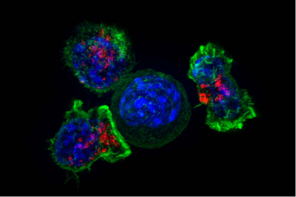
CAR-T therapy is a type of immunotherapy that takes advantage of the cell killing capabilities of cytotoxic T cells. This particular type of T cell is an important part of the human immune system which for many years, researchers have hoped might be useful in treating cancer.

Cytotoxic T cells surround a cancer cell
Cytotoxic T cells surround a cancer cell (taken from NIH)

The problem is, cancer cells are derived from your own cells so your immune system has no reason to target them for destruction. That’s where CAR-T therapy comes in. In CAR-T therapy, the T cells of the patient are taken from the blood and genetically modified so they express a chimeric antigen receptor (CAR). A CAR is basically a signalling molecule on the surface of the T cells that allow them to recognise cancer cells as worthy of destruction. These genetically modified T cells are then put back into the patient’s blood to target and kill cancer cells.

Because we know that different types of cancer typically have different types of signalling molecule on their surface, we can then use different types of CARs to help draw the cell killing T cells to the cancer cells. For example – nearly all types of B cell acute lymphoblastic leukaemia (ALL) have a protein called CD19 on the surface of cells so researchers have been working on CARs that specifically target cells which have CD19 on them.

B cell lymphoma cell is recognised by a T cell which has a CAR protein expressed on it
Modified from here.

Yescarta

The specific therapy that NICE has reported on this week is one called Yescarta from a company called Kite Pharma. Yescarta consists of taking T cells from the patient then using a system hijacked from retroviruses to genetically modify the cells. The genetically modified cells are then reintroduced to the patient in a single dose.

Clinical Trials of Yescarta have been initially promising. A phase II single-arm trial found that of 101 patients treated with Yescarta, 54% had a complete response and 52% survived over 18 months. All of these patients had refractory large B-cell lymphoma – that means they had a type of blood cancer that had already failed treatment and the patients had an average expected life expectancy of 3-4 months. While the trial showed that all patients had significant adverse events related to Yescarta, only 3% of patients died from the treatment itself.

Yescarta logo

Based largely on this trial the FDA approved Yescarta for certain types of lymphoma late last year and the European Commission granted Marketing Authorisation for the treatment this month.

Why not recommend it?

But NICE had some concerns. Firstly, they were concerned that the clinical trials for Yescarta, while promising, were insufficient to allow recommendation of the drug. While this particular type of lymphoma doesn’t have a standard therapy, yet, most patients are treated with scavenger chemotherapy and the Yescarta clinical trials haven’t proven that Yescarta is better than scavenger chemotherapy. The two therapies haven’t been compared side by side. This is quite a significant concern because, without side by side comparison, we can’t be sure that diverting patients from one treatment type to the other is actually beneficial for the health and life expectancy of those patients.

NICE aren’t saying no to Yescarta, they’re saying “not yet”.

The second concern NICE has is that the treatment is currently very expensive. The cost to the UK has not been made available, but in the US the treatment can cost $373,000 per patient. And this is something that NICE has to weigh up in its cost-benefit analysis. So far, Yescarta has been shown to prolong life by approximately 14 months in around 50% of patients. Is it justifiable to divert so many funds to a treatment that only works 50% of the time and we can only say extends life by a year (so far)?

A woman standing at a laboratory bench facing away from the camera and wearing a lab coat

The company that makes Kite Pharma was bought by Gilead Sciences last year, just two months before Yescarta was approved by the FDA. Last year Gilead Sciences had a revenue of $30.4 billion and a free cash flow of $15.9 billion. Gilead Sciences spent $12 billion in order to acquire Kite Pharma and its marketable therapy Yescarta. NICE perhaps has reason to be cautious.

Any other concerns?

If Yescarta were approved for the use in the UK, it would be the first of its type to be approved. There are potential concerns we need to consider when introducing both genetically modified cells into patients and playing around with the immune system in patients. In his article for McGill, Jonathan Jarry pointed out that “Autoimmunity is when our immune system improperly responds to something that belongs to us” and in the case of Yescarta we’re actively training the T cells to recognise cancer cells derived from our own cells. In fact CD19 isn’t just expressed on cancerous B cells, it’s found on all B cells – even the healthy ones. In the case of terminal cancer this might be a price worth paying but it has to be taken into consideration. Sudden activation of T cells in the body is such a strong response that patients can get very sick and even die from a condition called cytotoxic release syndrome.

We must also consider the reproducibility of the initially promising clinical trials. There is evidence that when repeated, less than 50% of clinical trial results are reproducible and many negative clinical trials go entirely unpublished which means a high likelihood of false positives, it seems prudent to take care when making decisions based on single clinical trials.

Conclusion

NICE - national institute for health and clinical excellence logo

While many patients were hoping for the approval of Yescarta by NICE, the limitations have proved too high at this stage. I don’t think this decision should be used to criticise NICE or the NHS, rather to encourage further research using reliable controls and a focus on bringing down the prices of promising new treatments. It also highlights an example where the novelty of new treatments can overtake scientific sense and lead to approving treatments which might currently be lacking sufficient clinical evidence supporting their use.

Meanwhile, the NHS will not withdraw funding for any patients already undergoing Yescarta therapy and will reconsider their decision given further research or reduced costs.


Cool science: Kick and Kill to cure HIV – an update.

A little while ago I wrote about an exciting new potential for HIV treatment. The cool idea was looking to solve a problem we haven’t yet figured out how to solve when it comes to treating HIV infection. You see, HIV is particularly tricky to treat because it forms what we call latent viral reservoirs. These are a group of cells that are infected with the virus but within which the virus isn’t ‘active’.

Antiretroviral drugs are really great, but they only target ‘active’ virus which means these reservoirs are essentially hidden away and protected. This is why people living with HIV need to stay on treatment for their entire lives.

So, researchers came up with a great idea – what if they could ‘kick’ the virus in the reservoirs into action and then ‘kill’ the now active virus using standard therapy. This scientific basis behind this ‘kick and kill’ approach made researchers think that this might hold the key to curing HIV and allowing people living with HIV to eventually come off their medication. Early studies were really promising and there was good evidence that this might be a useful approach to trial in patients.

So what happened next?

RIVER

The Research In Viral Eradication of HIV Reservoirs (or RIVER) study began in 2015 and concluded this year. This study was a randomised control trial (RCT) where 60 people recently diagnosed with HIV in trial centres across London and Brighton were split into two groups. One group received standard therapy – antiretroviral therapy (ART) only – this group acted as a ‘control’ group that could be used to compare the test group against. The test group were given a test therapy which consisted of four steps:

  • Patients were treated with standard ART until the ‘active’ virus was undetectable in the blood
  • Patients were then given a vaccine which would train or ‘prime’ the immune system to recognise the HIV virus
  • Patients were treated with Vorinostat which would ‘activate’ the inactive virus in the reservoir cells – this step is the ‘kick’ part of the process
  • The ‘primed’ immune system would then be able to ‘kill’ the newly activated virus

A four step diagram outlining the four steps included in the RIVER study - Step 1, ART is used to make sure HIV is undetectable. Step 2, two vaccines train the immune system to recognise cells which will be activated. Step 3, Vorinostat is used to wake the sleeping cells. Step 4, the immune system boosted by the vaccine attacks and kills the newly activated cells

What did RIVER find?

The results were surprising and unexpected. RIVER found that there was no significant difference in the size of the latent HIV reservoir between standard treatment (control) and ‘kick and kill’ treated (test) patients. It looked like ‘kick and kill’ was no better than standard therapy.

Even more surprisingly, each of the components –the standard antiretroviral medication, the vaccine which primed the immune cells and the Vorinostat which ‘kicked’ the inactive virus into action – all worked exactly as they should. But combining the multiple components wasn’t any better than ART alone.

Chief Investigator on this study, Prof. Sarah Fidler of Imperial College London said “In the RIVER study, we found that all the separate parts of the kick and kill approach worked as expected and were safe. The vaccine worked on the immune system, the kick drug behaved as we expected it to, and the ART worked in suppressing viral load in the body, but the study has shown that this particular set of treatments together didn’t add up to a potential cure for HIV, based on what we’ve seen so far.”

Does this mean we should drop the kick and kill approach?

Well, no, not really. Because this was just one iteration of the kick and kill approach using a combination of one type of medication and two types of vaccine. Researchers on the RIVER study aren’t really sure why it didn’t work as planned but until we understand the answer to that question it might still be a useful avenue for research.

a drawing of an HIV viral particle surrounded by blood cells and coloured with dark purple and brighter patches of fushia

The co-principal investigator and scientific lead from the University of Oxford, Prof. John Frater says “It is possible that the combinations of drugs we used weren’t quite right, but for this first study we didn’t want to compromise on safety by using stronger agents that might work better but could cause toxicity to the participants. It is possible that vorinostat was not quite potent enough to wake up as much HIV as was needed for the newly trained immune system to recognise. Equally, it is possible that a different sort of immune response to the one we induced is needed to target the HIV reservoir. All of these possibilities need to be teased out and considered to guide our next move in searching for an HIV cure.”

So what next? Collaborative research.

This RCT is an important step in investigating the possibility of curing HIV infection. Professor Abdel Babiker of the MRC Clinical Trials Unit at UCL, said “Although the results are disappointing, they are unambiguous because of the randomisation and completeness of follow up assessments. Because ART is so effective at reducing viral load, without the randomised control group of participants taking ART alone to compare against, we couldn’t have been so confident in knowing whether the kick and kill drugs had made any impact. It’s important that future HIV cure trials follow this approach and compare their outcomes to an ART-only group.”

A woman standing at a laboratory bench facing away from the camera and wearing a lab coat

Scientific experimentation using RCTs allows us to be confident in the findings – even if those findings are not as promising as we hoped they would be.

This particular trial was part of a UK collaboration group called CHERUB but there are collaborative groups like this all over the world. The value of collaboration allows experts from different backgrounds, with different expertise to come together and conduct research with wide ranges of participants. The importance of participant engagement in particular was praised by Prof. Fidler who said “They are not just volunteers, they are active advocates for support and they push us to go further all the time. They are helping to define where this research can go next and they are the real pioneers of new treatments.”


Find out more about this trial here and here.


Is Cannabis a Cure-all?

You might have seen in the media lately a lot of coverage about the use of cannabis to treat epilepsy and other conditions. It all started with the case of a young boy with severe epilepsy who was using cannabis oil to manage his seizures, with apparently great effect. Until, that is, his mother was unable to bring his treatment into the UK and his medication was seized at the airport.

These stories raise three questions:

  • What does UK law regulate in the case of medications based on cannabis
  • What is a cannabis oil?
  • Can cannabis treat any medical condition?

UK Law

In the UK, cannabis is a Class B drug – you aren’t allowed to possess or supply it and doing so can result in jail-time. This is a regulation under the Misuse of Drugs Act 1971, however cannabis is also regulated by The Misuse of Drugs Regulations 2001 which control the therapeutic use of drugs. Under this legislation, cannabis is regulated as a Schedule 1 drug which means it is not available for medical purposes and possession and supply are prohibited unless the Home Office approves.

cannabis plant seedlings

Cannabis: the sum of its parts

Cannabis refers to a group of plants which produce compounds called cannabinoids. Cannabis plants contain 113 different cannabinoids – so what exactly are we talking about when we talk about cannabis oil?

The two important cannabinoids to consider are tetrahydrocannabinol (THC) and cannabidiol (CBD). THC is the main part of cannabis that gives its psychoactive effects. It’s the compound that will make you feel ‘high’ if you smoke marijuana although this response is mediated by other cannabinoids too. It also stimulates release of the hunger hormone, ghrelin, which explains why people have an increased appetite when they take cannabis. It does this by binding to a specific receptor on the surface of cells in the brain.

CBD is non-psychotropic and it acts in a very different way to THC. But it might also enhance THC activity by increasing the number of receptors available for THC to bind to. It might also increase the levels of those natural endocannabinoids in the body.

In the UK, CBD is legal which means cannabis oils containing only CBD are legally available whereas THC is not legal.

four brown glass unlabelled bottles containing oil

Can cannabis treat disease?

In the UK, there are already two cannabinoid based treatments licensed for prescription. Nabilone is used to treat nausea and vomiting in people undergoing chemotherapy. There are other conditions it has been indicated for including IBS, fibromyalgia, chronic pain and parkinson’s disease however in the UK it is only permitted to help treat the side effects of chemotherapy.

The second cannabinoid based treatment available in the UK is Sativex which is used to treat the symptoms of multiple sclerosis including neuropathic pain and spasticity. Sativex is a cannabis extract which contains both THC and CBD.

Cannabis for epilepsy

When it comes to epilepsy – there is considerable evidence that THC can control convulsions through regulation of neuronal excitability and inflammation. But because it can make you high – it’s not an ideal avenue for therapeutic exploration.

Research into CBD for treating epilepsy is relatively new but initially promising at least for certain types of epilepsy and there is a drug awaiting FDA approval which can treat Lennox-Gastaut syndrome and Dravet syndrome, two severe forms of epilepsy. But this might not be sufficient in all cases – some patients may require different mixtures of THC and CBD to see an effect.

Cannabis for cancer

Cannabis can be useful in managing cancer-associated side effects in patients. It can act as both a pain reliever and a way to reduce nausea and enhance appetite. But there is early research that it might also kill cancer cells and stop them growing. In these cases researchers have looked at highly purified THC and CBD. Some trials have shown that combining chemotherapy with cannabis might have some promise. However, we have insufficient evidence to support its use as a cancer treatment either due to small study sizes or the research predominantly taking place in cells in the lab which is just not a good representation of what would happen in humans. We don’t know which types of cannabinoids are most useful, what doses are needed, what types of cancer respond, how to take them effectively and whether they should or shouldn’t be combined with other treatments.

Cannabis research

If cannabis is so promising, why don’t we do more research on it to bring it to clinical trial? Cannabis is a Schedule 1 regulated drug, it can only be used in research with Home Office approval. Schedule 1 drugs are so classified because they are not deemed to have medical usefulness. But researchers like Professor David Nutt are concerned that the medical usefulness of cannabis cannot be proven if research is prohibited.

Importantly, the recent media interest in cannabis use as a medical treatment has been useful in encouraging a UK government review on the therapeutic value of medications based on cannabis. This review will be undertaken by the Advisory Council for the Misuse of Drugs and may lead to a change in the legal status of cannabis and cannabinoids with regards to their use in medicine.

A woman standing at a laboratory bench facing away from the camera and wearing a lab coat

Alternative medicine

In the meantime, it is important to remember that while cannabis holds some promise as a potential therapy for many conditions, it is crucial to always follow professional medical advice when considering medical treatments. The research supporting cannabis use is limited and there are many questions about safety and efficacy that remain unanswered. For many conditions that cannabis might be useful for, we already have good medical treatments that can be used before considering an as-yet, unproven treatment. Cannabis oils are poorly regulated and might have wildly variable levels of cannabinoids and may even contain ingredients that are harmful. It is never advisable to buy medical treatments online or take medical advice from someone other than a qualified medical professional.


I talked more about this on my podcast, Skeptics with a K – on this episode. You can follow me on Twitter @AliceEmmaLouise for more.

Is sunscreen bad for you?

The weather has been glorious here in the UK, which means out come all the warnings to apply sunscreen copiously and frequently. It also means out come all the warnings that chemicals in sunscreen are dangerous.

But what does the science say?

Types of sunscreen

There are two main types of sunscreen: chemical or mineral. Chemical sunscreens contain chemical UV filters such as octinoxate and oxybenzone and some have retinyl palmitate added to them. Mineral sunscreens contain mineral compounds like titanium dioxide and/or zinc oxide. Chemical sunscreens absorb UV light and convert it whereas mineral sunscreens are reflective and act as a physical barrier. This means mineral sunscreens are often thicker and have a less pleasant texture on the skin and they leave your skin a little ghostly.

image of a person's knee with white sunscreen and a hearth drawn into the sunscreen

Chemical sunscreen – The warnings

When we see warnings about the dangers of sunscreen it tends to be related to three things:

  • Does chemical sunscreen cause skin problems such as contact dermatitis?
  • Does chemical sunscreen cause cancer?
  • Does chemical sunscreen cause birth defects?

So what are these concerns based on?

Contact dermatitis

Some people have skin reactions to chemical sunscreens – this occurs in less 1% of users and can be a response to fragrances, preservatives or the UV absorber itself. Sensitivity can develop after using a particular formulation for a long time. If you have a sensitive reaction to sunscreen you can try switching formulations, or you can switch to mineral sunscreen which is less likely to cause a reaction. And of course, see your doctor if you’re worried.

Causing cancer

Some studies suggest that oxybenzone can cause hormonal changes in cells grown in the lab. These hormonal changes have been confirmed in animals like mice but have not been reliably shown to occur in humans. Hormone changes can cause cancer so some people believe that oxybenzone can cause cancer. To date this has not been shown to be the case. Oxybenzone has not been shown to cause the DNA mutations needed to cause cancer and hormonal changes are not always linked to cancer. This evidence is insufficient to prove any link between oxybenzone and cancer.

an image of a small white mouse standing on a white background

Retinyl palmitate is sometimes found in sunscreen. Retinyl palmitate is derived from retinol or vitamin A and it acts as an antioxidant. Retinol generates reactive oxygen species (ROS) when exposed to UV radiation and ROS are able to damage DNA. This is the basis for the concerns that Retinol will cause cancer. Studies in mice did not show that retinol combined with UV radiation causes cancer. There is no data published in humans to suggest that retinyl palmitate causes cancer.

A recent meta-analysis confirmed that there is no evidence supporting an increase in cancer risk caused by sunscreen use.

Causing birth defects

There is evidence that medicinal retinol pills can cause birth defects however this has not been shown to be the case with topical retinol application. Still, as a precautionary method it is advisable that pregnant women do not use a sunscreen containing retinols for the duration of their pregnancy.

The context

It is important to note that while there may be some evidence suggesting some level of risk associated with chemical sunscreen risk – this must be taken within the wider context.

Skin cancer

There are two main types of skin cancer – melanoma and non-melanoma skin cancer. Non-melanoma skin cancer includes basal cell carcinoma and squamous cell carcinoma and is largely treatable if it’s caught early. Non-melanoma cancers are the most common type of cancer. Melanoma skin cancer is an invasive form of cancer that is the 5th most common and at late stages is usually considered incurable. At early stages it is highly treatable but this form of cancer can progress rapidly and requires early intervention.  Both types of skin cancer are on the rise in the UK and this is linked to increasing sun and sunbed exposure. UV light exposure accounts for 86% of all melanoma cases, in the UK. Studies in Australia have shown a reduced rate of melanoma with regular sunscreen use.

A white sunhat with a black ribbon on a table with a pair of blue lensed sunglasses

Does sunscreen prevent cancer?

There is evidence that regular sunscreen use reduces pre-cancerous conditions and prevents skin cancer. However, the research into the efficacy of sunscreen is highly variable. This is partly because people are prone to using sunscreen in order to extend their time in the sun and misunderstand the most effective ways to use sunscreen. Chemical sunscreens should be applied to the skin 30 minutes before going into the sun and should be reapplied every two hours or more often if you are perspiring or swimming. Even waterproof sunscreen will be removed by towelling down after a swim. Sunscreen does prevent sunburn however research shows that people who only rely on sunscreen to protect themselves from UV damage burn more often than people who also practice sun avoidance habits. A person who has suffered sunburn more than twice in their life is twice as likely to get melanoma.

So what should you do?

While there is evidence that chemical sunscreens can have some detrimental effects on the body – the evidence is overwhelmingly clear that over-exposure to UV light causes skin cancer. Not only that, the research shows that the benefits of using sunscreen far outweigh the risks. Unless you are completely avoiding any UV light exposure then in my opinion, using sunscreen is a risk worth taking. In addition to wearing sunscreen and reapplying regularly, you should aim to avoid direct sunlight during the hottest hours of the day or wear clothing that covers your skin. And don’t forget, you might not burn through glass but you can still get UV skin damage through glass!

Extra reading:

https://www.skincancer.org/prevention/sun-protection/sunscreen/sunscreens-safe-and-effective

https://www.consumerreports.org/cro/sunscreens/buying-guide/index.htm

https://www.popsci.com/sunscreen-harmful#page-2


Autism Spectrum Disorder (ASD) and Aluminium

Recently, Professor Chris Exley from Keele University claimed that his research “provides the strongest indication yet that aluminium is a cause of ASD”. In an article originally published in the Hippocratic Post and reproduced by the Daily Mail, Exley claimed that brain tissue from five donors diagnosed with Autism Spectrum Disorder (ASD) contained “some of the highest values yet measured in human brain tissue” of aluminium. He postulated “perhaps we now have the link between vaccination and autism”.

This controversial claim comes almost two decades after Andrew Wakefield published the now discredited and retracted research into ASD and the measles, mumps and rubella (MMR) vaccine which was linked to a decline in vaccination rates and recent outbreaks of measles.

I spoke to Tom Chivers from Buzzfeed about my concerns with this paper. In his article you can read concerns from myself and eminent academics Professor Dorothy Bishop and Professor Jonathan Green, renowned experts in neurodevelopmental biology and child and adolescent psychiatry respectively.

Such a controversial claim which can affect the health of children worldwide really needs some solid supporting evidence. So, let’s take a look at the paper itself.

Aluminium in brain tissue in autism

The paper is due to be published in the Elsevier Journal of Trace Elements in Medicine and Biology in March 2018 and the accepted paper is available to read online. The authors introduce the study saying “we have measured aluminium in brain tissue in autism and identified the location of aluminium in these tissues”. The paper can be split into two sections.

Section one

The authors measured the levels of aluminium in brain tissue taken from five donors aged 15 to 50 who had all been diagnosed with ASD prior to their deaths. They took 0.3g tissue samples for four (occipital, frontal, temporal and parietal lobes) or five (they added hippocampal tissue where it was available) brain regions. They measured three replicates of each brain region using spectrometry.

The problems with the methods and data from this section are three-fold:

  1. The authors took samples from only five brains – all five donors had died with a diagnosis of ASD. There were no healthy control samples used anywhere in this study. This is a particular problem because without a healthy control, it is impossible to really say whether the aluminium levels measured in the brains of the donors is actually higher than that of a non-autistic brain. There is nothing to compare the measurements to.
  2. Importantly, the authors do not provide information on how the donors died. We have to take their word that the cause of death does not account for variation in brain levels of aluminium. An important part of the scientific publication framework is that this sort of information should be easily available to the reader. We need to be able to come to our own conclusions based on the data presented. That this information is missing is quite concerning.
  3. Finally, in some cases the three replicates from each brain region per donor did not corroborate each other. In one case there were three measurements of 0.01, 0.64, and 18.57μg/g. Similarly, another donor had measurements of 2.44, 1.66, and 22.11 and another, 1.71, 1.64, and 17.10μg/g. The authors averaged these data, however such an average does not represent any of the three measurements and is an inappropriate way to analyse these figures. In fact, such variation between measurements might even hint at an anomalous result, which requires further assessment to adequately rule out.

Section two

In the second section of this paper the authors took samples of brain tissue from the valuable Oxford Brain Bank. Again, they took tissue from a selection of donors who had been diagnosed with ASD before their death. Again, the authors failed to express how the donors died. The authors took samples from 10 donors aged 14 to 50 and stained them with a dye that specifically sticks to aluminium. The dye can then be visualised with a fluorescent microscope to see where in the brain tissue it can be seen.

Again, the issues with this section are three-fold

  1. This section also lacks the use of ‘healthy’ controls. The authors do show presence of aluminium in the tissue samples of their ASD donors but we have no indication of whether this is, in fact, more than we might see in a healthy brain.
  2. This section is also lacking a positive control – a sample that has a known amount of aluminium in it. This is important for interpreting the staining of the dye; if the dye is very sensitive then we might see loads of staining for just a small amount of aluminium. We need to compare it to a known sample.
  3. Later in their interpretation of these data the authors discuss the presence of aluminium in specific cell-types which they surmise might be lymphocytes. However, they do not use any detectable markers to propose this, they base it only on how the cells look. Cell appearance (also known as morphology) is a useful initial indicator of cell-type but it is important to confirm this using specific markers for those cells.
1-s2.0-S0946672X17308763-gr1
A figure taken from the paper which apparently shows the accumulation of aluminium in specific cell types

Overall the data from this paper is insufficient to draw the conclusions made in the Hippocratic Post and The Daily Mail and it is my concern that such reporting is irresponsible. While the two articles conclude that aluminium present in the brains of people with ASD might be caused by vaccination, there is no investigation whatever into the source of such aluminium in this study. There are no data presented in this paper sufficient to implicate vaccination in these findings. To draw the link between aluminium in the brain of people with ASD and vaccinations makes for a striking headline but the data do not support this claim and it could therefore be viewed as scare-mongering.

I am surprised that this paper was published in the Journal of Trace Elements in Medicine and Biology because the lack of control data makes the findings inconclusive. I am yet more surprised that this inconclusive data has been reported in such a paper as The Daily Mail, when such media attention can lead to parents making potentially dangerous decisions for the health of their children.

Please let me know below what you think about the reporting of this science?

How many mutations does it take to create a cancer?

Last week I wrote a post all about how we understand cancer to arise. I explained how mutations in certain genes for certain proteins can drive cancer and I explained some of the important families of proteins such as oncogenes and tumour suppressors.

But even that complicated post was an over-simplification. Sure, a single mutation in a single gene for a single protein might cause cancer, but actually we know that many cancers have several mutations. Sometimes, several hundred mutations.

Several hundred mutations

many mutations per cancer
Image adapted from this paper. A graph showing a number of non-synonymous mutations per tumour against tumour types ranging from colorectal cancer to glioblastoma. (See the text for some example figures).

The above is an image which shows just a few examples of some cancer types and the number of mutations you might expect to find in them. Recognise the word non-synonymous from last week? Non-synonymous mutations are those which cause a change in a protein. Whereas synonymous mutations are effectively “silent” (they don’t change a protein) so we tend to focus on non-synonymous ones when it comes to disease. You can read more about them here.

From the image you can see that some types of cancers can have several hundred mutations per tumour – colorectal cancer for example can have between 500 and 1200 mutations. Lung cancer can have between 100 and 200 (interestingly this is much lower for lung cancer in the “never-smoked” category which has a considerably smaller number of 25) and breast cancer between 25 and 100 mutations per tumour.

The problem

What this means is that when it comes to understanding what drives cancer we need to know how many of these mutations are actually relevant to the disease. Do they all contribute to “driving” cancer or are some of them “passenger” mutations? If only a few contribute, then how many?

Often, we treat cancer by designing drugs which target specific proteins. If we suspect a cancer is “driven” by an overactive protein then we can treat patients with an inhibitor of that protein – something that blocks the protein’s function. But for us to do this, we need to know which mutation, potentially hundreds, is actually relevant. It’s like trying to find a needle in a haystack.

The study

The paper published in October 2017 in Cell is an analysis of 7,664 tumours across 29 different tumour types. They looked at all of the mutations each of those tumours had and focussed on the non-synonymous ones, the ones that actually change the proteins inside the cell. But they knew that even with protein changes, not all of the mutations present actually cause cancer. They wanted to establish how many might be responsible for the disease.

“Normal” cells acquire mutations all of the time but this happens at random so sometimes it will affect proteins and sometimes it won’t –  we get a reliable mixture of non-synonymous and synonymous mutations that are all mostly harmless even if there is a change in a protein. This is useful information because we can establish an “expected” ratio between the two. In cancer cells, the mutations that change proteins and allow a cell to survive and grow more than it should (i.e. the mutations that drive cancer) are passed on to more cells as the cancer cells replicate. This means you get a skewed ratio in cancer cells – more non-synonymous mutations than synonymous ones. By figuring out how many more mutations there are than we expect from the “normal” cells we know that those are the ones allowing cells to replicate in an uncontrolled way – therefore those are the ones that are driving the cancer.

And that’s exactly what the authors of this study did.

What they found

What they found was that although some cancer cells have hundreds of mutations, only a few of those actually drive the cancer. The average number of cancer drivers per cancer type is just four and the numbers only range from one to ten. So, we can narrow our focus to between one and ten mutations per tumour. We just need to figure out which ones.

This is phenomenally useful information because we can then build further on this knowledge to understand which mutations are the common driver mutations and focus our drug development on those specific mutations. We have a significantly smaller haystack and we can start to really get very specific with our cancer therapies! The better our understanding of how cancer really works, the more complete our understanding of all of the complicated processes involved, the better able we are to design clever therapies that really benefit patients.

What drives cancer?

When it comes to understanding cancer, getting our heads around the complicated process of how it forms in the first place is vital. When we know what drives cancer then we can figure out how to diagnose it and ultimately, how to treat it.

So far, research has shown us that the vast majority of cancers are caused by some level of genetic mutation. This is something we’ve known for a long time and something we use in order to treat different types of cancer. For example, a reasonable proportion of breast cancer patients have a mutation in the gene for a particular protein called HER2 making this protein more abundant than it should be and allowing cancer cells to grow and grow. So scientists have designed a therapy that blocks, or inhibits, HER2 – a drug called Herceptin. The cancer cells have been relying on HER2 for their growth, they’ve becomes entirely dependent on it which means when we treat patients with Herceptin, HER2 activity is reduced and the cancer cells die.

Another thing we know about cancer, is that it’s hugely genetically unstable. This means cancer cells acquire loads and loads (sometimes many hundreds) of new mutations as the disease progresses. What is important is to understand which of these many mutations are actually fundamental to the survival of the cancer. Which ones are truly driving the disease, allowing it to survive and progress and spread?

This can be particularly complicated for us to understand because a mutation in one gene can do lots of different things.

Genes

First things first, let’s define what a gene is. When it comes to DNA we can think of an organism’s entire genome like a recipe book. The entire human genome book allows our cells to work together to cook an entire human. If we were cooking a really complicated meal like Christmas dinner we would need to cook all the different components and bring them all together to make the meal. You have to cook the main dish and all the side dishes, your carrots and parsnips and brussel sprouts, maybe two kinds of potatoes and a soup for the starter. Every person’s Christmas dinner might look slightly different but we all agree it’s a complicated meal and there are lots of components. The human body is also pretty complicated and is made up of components. A human body is made up of organs, which at the most basic level are made up of cells. Your cells are kind of like the ingredients for each dish in your Christmas dinner. Your organs are kind of like those individual dishes. They all come together to make the whole human. So, in order to make such a complicated thing, we need lots and lots of recipes. Within the human genome we have 23 pairs of chromosomes, which we might think of as chapters of the recipe book. Within each chapter are individual recipes, or genes, which allow cells to make for individual building blocks (ok, ok, I’m mixing my metaphors…).

The basic building blocks in the human body are cells. Cells come together to make our organs, and organs come together to make a human. But our cells need to do specific jobs depending on what kind of cell they are; they need to communicate with one another within the organs they are a part of. This is what makes our heart cells contract all together to pump blood around the body or it allows all of our lung cells to know that they’re supposed to be lung cells. This communication is done through proteins, which can pass signals along complicated pathways transmitting the message from protein to protein and giving each cell instructions on how to function. A single gene is a recipe for an individual protein. Each gene is made up of many thousands (depending on the protein size) of what we call bases which are signified by the letters A, T, C and G. There is an extra layer of complexity as the code that makes up a single gene is organised into groups of three letters each group referred to as a codon. Each codon codes for a single amino acid and these amino acids build up in long sequences to eventually form proteins.

DNA to protein
In order to convert the genetic code the cell must convert DNA into an intermediary molecule called RNA. This can then be translated into protein. Each ‘codon’ (three letters) is a code for one amino acid which build up in sequences to make proteins.

A mutation is change in this code. And that tiny change can have a number of outcomes.

Ultimately – the ones we’re worried about are those that change how particular proteins behave.

How do protein mutations drive cancer?

On a very basic level, proteins basically control everything. They regulate which cells go where, how big cells grow, how much cells replicate and how cells function within their organs. Some proteins are crucial to very specific pathways related to cell survival and cell replication (referred to as proliferation). These proteins we refer to as oncogenes or tumour suppressors. They carefully regulate how cells grow and survive. If these pathways become deregulated, cells can grow out of control and survive when they shouldn’t. This is how cancer forms. These pathways, and therefore these particular proteins, are fundamental in driving cancer. Many of the most commonly mutated proteins in cancer are either oncogenes or tumour suppressors. But what exactly are these proteins?

An oncogene is a protein which promotes cell growth and cell survival. In normal cells these proteins are carefully regulated. They become activated and they pass on the message to tell the cell to grow or survive and then they get switched off until they are next required. Oncogenes can be mutated in such a way that their activity is increased – either there are loads of copies of them all passing on the “grow and survive” message at once, or they are unable to be switched off and they are constantly telling the cell to grow and survive. In this way, a mutation in an oncogene can drive cancer.

A tumour suppressor is one of those proteins that helps regulate the growth and survival pathways. Tumour suppressors can switch oncogenes off, or they can oppose them by sending the opposite message. They serve to slow down the “grow and survive” message and keep our cell growth and survival finely tuned. In this way, when functioning properly, tumour suppressors prevent cancer. A mutation that blocks these proteins from doing their job will therefore promote cancer.

Oncogenes
This is adapted from my PhD thesis. Here are two pathways with the various proteins that ‘transmit’ the signal through a cascade of activation. One pathway leads to cell replication (proliferation) and the other to cell survival. Some of these proteins are particularly crucial in promoting these pathways – PI3K and Ras, for example, are oncogenes. Some of these proteins are crucial in regulating or slowing down these pathways – PTEN, for example, is a tumour suppressor.

Different types of mutation

This complexity of the genetic code means there are number of different ways in which a gene can be mutated.

We can split these into two broad groups – synonymous and non-synonymous. Synonymous mutations are ones where the mutation doesn’t lead to change in the sequence of amino acids in a particular protein. They are effectively silent mutations because they don’t change proteins.

But many gene mutations are non-synonymous; one way or another, they cause a change in the protein produced by that gene.

Of these we can have:

Missense mutation is a mutation where one amino acid is switched out for another. The importance of that particular amino acid to that particular protein designates whether or not this change has a big impact on the protein function. It can increase activity of an oncogene, or it can prevent an active oncogene from being switched off. Alternatively, it can block the action of a tumour suppressor.

Nonsense mutation is a mutation which leads to termination of the protein production at that position in the code, leading to a shortened or truncated version of the protein which may severely alter the ability of that protein to function normally. A truncated tumour suppressor, for example, isn’t able to switch off cell growth and might promote cancer.

Insertions/deletions occur when a few letters are added to or removed from the code which can completely alter the entire remaining sequence leading again to protein truncation. Large deletions can remove entire genes preventing the production of that entire protein. This can include duplication mutations when a particular region of DNA is duplicated which may enhance or reduce the activity of a protein.

Large-scale mutations in chromosomal structure – these can be very complicated including the amplification of entire genes giving you far too many copies of a particular protein. They can also create entirely new proteins by fusing two separate genes together.

What does all this mean?

Hopefully it’s clear that the variety of mutations and proteins involved in driving cancers is quite complicated and that makes understanding cancer difficult. However, we might also see opportunities for using knowledge to treat cancers. If we can establish how frequently a particular cancer is cause by an overactive oncogene then we can start to find treatments that block or inhibit those oncogenes. These are strategies that have led us to discover drugs like Herceptin, which blocks the oncogenic HER2 receptor I talked about at the beginning of this post. But it also means we need to fully understand which mutations drive cancer in order to establish treatments for them.

Next week I’m going to talk about a particular study that looked at these driver mutations that was published just this month. The researchers wanted to get a more complete picture of how many proteins we might need to target in order to effectively treat specific types of cancer and what they found was really interesting.

For now, I’d love some feedback on this post because it’s quite science heavy – if there’s anything I could have explained better or in more detail then let me know in the comments and maybe I can write more on these topics in the future.


*edited to correct some of the HER2 science – thanks to my good friend Dr Jenny Smith-Wymant for pointing out my mistake.

Will we ever cure HIV infection?

So far on this blog, I’ve talked a lot about cancer. I’ve been working in cancer cell biology for a number of years, and it’s an area that a lot of people are interested in, so it makes for a sensible topic. By training, though, I am a cellular and molecular physiologist. I’m interested in how all cells work; they don’t have to be cancer cells.

The human body, and cells in particular, amaze me. I love learning the intricate details of exactly how our cells work on a molecular level, but I love how we can identify little changes that might be useful in understanding disease and treatment even more.

Earlier this year I came across an interesting study on treating individuals infected with the Human Immunodeficiency Virus (HIV). HIV is a complicated virus and has long been a source of fear. But by understanding the science of the virus we can look to treat infection and perhaps dispel some of those fears. So here’s a bit about HIV, and then a bit about a cool study.

HIV

HIV is a type of virus that we call a retrovirus. A retrovirus carries its genetic material as a single strand of RNA, instead of the double-strand of DNA we use as humans. To transmit its genetic material the virus needs to construct DNA, which it does inside a host cell. In the case of HIV, the host cells are cells within the human immune system – white blood cells called helper T cells.  Once an HIV molecule (a virion) gets inside a human host cell, it uses an enzyme within the virus to transcribe a piece of its RNA into DNA. It then slots that DNA into the host cell genome. It can remain dormant for many years but eventually the host cell machinery produces many, many copies of this piece of DNA, releasing more HIV virions into the blood stream. Essentially, the virus hijacks the host cell’s normal processes in order to make copies of itself.

Picture1
The life cycle of HIV: the virus enters a host cell and releases its RNA which is transcribed into viral DNA. Viral DNA is integrated into the host cell DNA where it can lie dormant for many years. Upon activation, the host cell produces viral RNA allowing the generation of new virions which are released into the blood and are able to infect new host cells.  Image taken from Encyclopaedia Britannica.

The HIV virus does not need all the machinery required for its own replication therefore the HIV genome is tiny, consisting of only nine genes. By comparison, the human genome consists of around 20,000 genes!

Treating HIV

These days we are pretty good at treating and managing HIV infection. HIV infection isn’t currently curable, but we’re so good at treating it that many patients live a long, healthy life while carrying HIV. There are many caveats to that, including political problems related to getting treatment to all the parts of the world that need it.

The very first treatment we produced for HIV was an inhibitor that blocked the action of a protein important in converting the virus’ RNA into DNA, so preventing its subsequent replication and spread throughout the body. Since then we’ve designed different types of antiretroviral therapy (ART) which target different processes within the virus’ lifecycle. We can stop the virus getting into a cell, stop its RNA converting to DNA, stop that DNA integrating into the human genome and stop the virus from assembling and breaking out of the host cell. The problem is, once the virus is integrated into the host cell DNA it can lay dormant, untouched by inhibitors even when the viral load is reduced in the patients’ blood. We have to maintain treatment to prevent this dormant DNA from activating and the virus spreading around the human body. It is possible, too, for the virus become resistant to each therapy meaning careful assessment of the patient and modification of the therapy accordingly. In this way, we can manage the infection long-term. But what if we could figure out a way to target the dormant virus sitting in host cell DNA?

New study

Picture2
Title and authors of the new study published in September 2017 in PLOS Pathogens

A new study published in September aimed to do exactly this. Earlier research had shown that a particular type of drug can ‘activate’ the host-cells which hold dormant HIV DNA. These ‘activated’ cells produce lots and lots of RNA for the virus which “kicks” the hiding virus back into action. Using this principle, the standard anti-retroviral drugs are then able to “kill” the virus specifically targeting this pool of dormant virus that standard therapy alone is unable to target. This strategy is referred to as “kick and kill”. While other researchers have studied this principle before, the effect on host-cell activation has been minimal which means only a small proportion of cells holding dormant HIV can be targeted. The larger this proportion, the closer we can get to activating and killing every cell holding dormant-HIV and the closer we can get to curing HIV infection.

In this study the authors used a synthetic compound, SUW133, which would they hoped might activate more host cells and therefore be more successful.  Their initial work looked at the effectiveness of the compound in cells taken from six patients. They showed that control treated cells (treated with only media) had relatively few RNA molecules from the HIV virus whereas when they took those same patient-derived cells and treated them with the SUW133 the number of viral RNA molecules increased significantly. This indicates that SUW133 does, indeed, activate the dormant virus and kicks the host cells into action.

Picture3
Data taken from the paper. Comparison of control treated human patient cells with SUW133 treated human patient cells. Samples derived from the same individual patients are connected with a line showing the increase in activation of cells per patient when treated with SUW133.

They then wanted to look at what would happen in a living organism, so the researchers had to infect humanised mice (mice genetically modified to express human genes) with HIV. They did this for 3-4 weeks, after which they treated the mice with antiretroviral therapy for 2-5 weeks. Once they were sure that only dormant HIV would remain in the mice, they treated them with their special synthetic compound and looked in the blood for evidence of HIV-host-cell activation. In mice treated with a control (inert) compound they found relatively low proportions of specific cell types (CD3+/CD4+ and CD3+/CD4-) that expressed a marker for activation (CD69) however upon treatment with the active compound (SUW133) the proportion of “activated” CD69 positive cells rose dramatically.

Picture4
Data from the paper. The proportion of  activated (CD69+) blood cells in the blood of HIV positive mice is much higher in mice treated with SUW133 compared to control treated mice.

What’s more, the researchers showed that a small proportion (around 25%) of these activated cells were dead or dying which shows promise for the additional treatment with standard antiretroviral therapy to fully “kill” the virus.

Overall the authors have shown that they can activate dormant HIV which they hope will give us the opportunity to kill this currently untreatable pool of the virus and one day give us a cure to HIV infection.

What’s clear is that this is still early days – the researchers need to fully establish the effect of this “kick” strategy when combined with the “kill” treatment (ART). However, it is incredibly promising that we might someday be able to activate host-cells carrying dormant HIV genetic material so that we can remove those cells entirely. If we are successful, we could go from long-term HIV infection management to HIV-infection cure.


Science or Pseudoscience: Ancient Chinese Calligraphy Ink and Cancer Treatment

When it comes to reading about cancer cures in the media it is usually good advice to be pretty skeptical. Sometimes the story lends well to skepticism – like this one recently covered in a few outlets where it was proclaimed that scientists had found a cure for cancer in ancient Chinese calligraphy ink. It’s a story that has everything – including lasers. Here is a romantic idea that the cure for cancer might lie in an ink that is produced from plants and has been handed down between generations as a way to share writing and art. The proposed treatment is billed as non-invasive and specific only for cancer cells – could this really be science?

I was particularly suspicious when I saw that the representation of the story on Natural News, a prominent proponent of pseudoscience, looked a lot like the story on Science Alert which is typically more of a reliable source on science stories. So I did some digging into the data for this finding.

The paper that was covered in the media is titled “New Application of Old Material: Chinese Traditional Ink for Photothermal Therapy of Metastatic Lymph Nodes” and was published in the journal ACS Omega in August 2017. The researchers were based in Shanghai distributed across various institutes at different hospitals or universities. They had been working on a relatively new cancer therapy called Photo-thermal Therapy (PTT).

Photo-thermal therapy (PTT)

In cancer treatment, PTT is the use of specific types of nanoparticles to generate cell damaging heat specifically at the tumour sites. Scientists take a material that can be stimulated with light – typically infra-red – causing the generation of heat and subsequent tumour cell death. The problem is that many of these nanoparticles are toxic or expensive so in order to make the treatment as efficient and effective as possible, we need to find just the right material.

Hu-Kaiwen

Hu-Kaiwen (Hu-ink) is an ink that has been used by calligraphers in China for hundreds of years. It’s derived from plants, is mostly made up of carbon and it is black in colour which the materials used for PTT tend to be. So scientists got to wondering if it might be useful for PTT.

Firstly the authors of this study looked at the stability of the ink. They diluted it in different things including water and saline and made sure it was stable when stored over time. They looked at the structure of the ink and noticed that it typically forms small aggregates of 20-50nm in diameter – nanoparticles. They confirmed that the core component of the ink was carbon and they stimulated different concentrations of the diluted ink with infra-red lasers and tested the temperature. They found that Hu-ink was more efficient at converting light into heat than most other PTT materials reaching temperatures of 55°C after five minutes irradiation.

Hu-ink for PTT in cancer cells grown in the lab

In research we use cell models of cancer which we refer to as cancer cell lines. These are cells that have originally been taken from patients with different types of cancer but are grown and stored artificially in the lab. We use them so we can test things out on cells that behave like cancer but aren’t in a human body before we move on to doing tests in living organisms. The researchers in this study used some cancer cells originally derived from colon (SW-620) and colorectal (HCT-116) cancer. First they treated the cells with just the Hu-ink and the cells tolerated it really well proving that the ink solution itself wasn’t toxic. Then they treated the cells with the ink and combined it with irradiation with the infra-red laser.

Hu-ink
HCT-116 cells treated with Hu-ink and infra-red compared to HCT-116 cells treated with infra-red only. Image modified from the paper.

The image above is part of a figure taken from the paper itself. There are three images of HCT-116 colorectal cancer cells. In the top image, the cells were treated with only the laser for five minutes. They were not given any Hu-ink. In the middle image the cells were treated both with the Hu-ink at a medium dose for two hours and the laser for five minutes. In the bottom image the cells received a high dose of the Hu-ink for two hours and the laser for five minutes. The red cells are cells stained with a marker of cell death. The green cells are living cells. You can also see that the dead cells are much smaller than the living cells. In the top image where cells did not receive any ink – all of the cells are alive. In the bottom image where the cells had a high dose of ink – all of the cells are dead. In the middle image with a lower dose of the ink, there are some living cells and some dead cells. In other words – when you treat cancer cells with both Hu-ink and infra-red, the cancer cells die.

Hu-ink for PTT in mice

The researchers wanted to be sure that this technique was safe in living organisms and that it was able to kill cancer cells in living organisms. So they took mice with cancer of the lymph nodes and injected Hu-ink into the tumour which they then irradiated with infra-red. After an allocated treatment time, they removed the tumours from the mice and measured them.

Hu-ink2
Lymph node tumours taken from mice treated with Hu-ink and infra-red versus control treatments. Image modified from the paper.

For this experiment they had three control conditions – NS (the tumours were injected with saline), NS plus laser (the tumours were injected with saline and treated with a laser) and Hu-ink (the cells were injected with Hu-ink). They had one test condition – the Hu-ink plus laser. The test condition is the only one that the authors predicted would have an effect on tumour size. And that’s exactly what they saw. Lymph node tumours treated with Hu-ink and infra-red were significantly smaller than the control conditions. They also saw that the surrounding tissue wasn’t damaged suggesting that the treatment is safe.

Study conclusion

This study is a proof-of-principle study. The authors have shown that this ink can be used in PTT therapy with a positive effect and in a safe way in mice and in the lab. It is a very small scale study and it is a single study. It needs replicating before we can be confident in the result and it needs to be studied on a larger scale and have many more safety tests before we could begin to think about using it in patients. But it is a really promising study. It takes a treatment we already use and aims to make that treatment safer and cheaper and more available to patients. It is a non-invasive treatment and it can be used in ways that reduces tissue damage to healthy tissue while targeting cancer cells for cell death. It’s a great example of some really cool science, using materials that have been available for many years and applying them to modern techniques.

It might seem far-fetched to say ancient ink can help us treat cancer, but really it’s just cool science!

Cool science: using Zika virus to treat cancer?

Throughout the last two years there has been a great deal of news on the Zika virus – a virus spread by mosquitoes which was first identified in 1947 in Uganda. In a normal healthy adult Zika fever causes relatively mild symptoms or even none at all however the 2015-2016 Zika epidemic gave rise to widespread concern due to its propensity to cause microcephaly and brain defects in babies infected with the virus during development. The epidemic was declared over in late 2016 although there are still travel warnings to certain areas where the mosquitoes known to carry the virus are prevalent.

But research into this particular virus highlighted an interesting trait that we might be able to take advantage of – Zika has a preference for stem cells.

Zika and stem cells

The reason Zika virus is particularly dangerous in developing babies is that the virus causes damage in stem cells in the brain. A stem cell is a cell that isn’t yet programmed. They’re really important in development because when you create a baby you start with one sperm cell, one egg cell and these cells needs to combine, proliferate and then differentiate into all the different types of cell within the human body. Stem cells are unique cells that can be programmed or ‘differentiated’ into all sorts of different types of cells. Once a stem cell has differentiated it can’t turn back into a stem cell – a differentiated cell is committed to only ever being that cell type. The adult body has very, very few undifferentiated cells but a developing foetus has plenty. This explains the risk of Zika infection during pregnancy as Zika has been shown to target neural progenitor cells – a type of undifferentiated cell in the brain – that might lead to the microcephaly seen in babies infected with the virus during their development.

480px-Zika-chain-colored
Crystal structure of the Zika virus

Zika and brain cancer

Cancer stem cells are quite a complicated thing that I’m not going to try and do justice in this post because it’s a topic that deserves its own post. Scientists believe that some cancers do have associated cancer stem cells. How exactly cancer stem cells might contribute to cancer progression is far from fully understood. However, we do believe that the presence of cancer stem cells might contribute to cancer therapy relapse. This is particularly concerning in glioblastoma – an aggressive form of brain cancer, which has poor survival rates despite our best efforts. Without treatment, median survival is around 3 months from diagnosis. With treatment we are able to extend that survival to 12-15 months however the cancer usually recurs. Scientists believe this recurrence is all down to the presence of cancer stem cells.

The study

So here’s the clever part. Cancer researchers know we have a problem in treating glioblastoma. They also realised that Zika virus is a relatively mild virus, which attacks stem cells. The adult brain doesn’t really have stem cells – unless the adult has glioblastoma. Cancer stem cells in the brain lead to cancer relapse; Zika attacks brain stem cells. Maybe we can make use of these two pieces of information.

So, scientists did some experiments. Firstly, they took some glioblastoma cells from patient tumours and they grew them in a dish in the lab. Then they infected them with Zika virus. They looked at either glioblastoma differentiated cells or glioblastoma stem cells. And they looked at the infection rate. Over 48 hours, over 60% of the stem cells were infected and this increased over time as the virus spread. The differentiated cells were infected too but not as much. What was especially interesting was that the stem cells infected with the virus had severely reduced ability to multiply and they had an increase in cell death. This was specific to only the stem cells and didn’t affect the differentiated cells. The virus kills cancer stem cells and prevents them from spreading.

Next the scientists took some patient tissue samples – this allowed them to look at a whole mixture of cells in a slightly more normal context without having to infect patients. They infected the tissue samples with Zika and saw that cancer samples were infected successfully and the virus only hit the stem cells and not the other cell types in the sample. They also looked at some brain samples from epilepsy patients and the virus didn’t infect them showing that the virus really is specific for stem cells!

jem.20171093
Glioblastoma tissue sample (from the paper)


Finally, they used the virus to treat mouse models of glioblastoma. They took mice with glioblastoma tumours in the brain and infected them with the virus. They saw that the tumours were much smaller and the mouse had improved survival when they were infected with the virus compared to control treated mice. They went on to show that they got an even better effect when combined with other glioblastoma treatments.

Benefits

Current treatments have two problems when it comes to glioblastoma. Firstly, the cancer stem cells make recurrence almost inevitable. This could drastically improve average survival times. Secondly, all brain cancer treatments have to be able to cross the blood-brain barrier in order to get through to the cancer cells. The blood brain barrier is an important way to keep things out of the brain where they might cause damage but it also serves as a way to keep brain tumours trapped in and harder to treat. Zika is great at crossing the blood-brain barrier.

What next?

We’re still such a long way from this being a useful patient treatment. In order to use this as a treatment we need to modify the virus in such a way that it will not spread from person to person and it will not cause the patient any harm. Currently virus work is always done in very specialised laboratories with expert training on how to prevent spread and with many, many precautions. If it were to be used as a therapy we’d need lots and lots of precautions to make sure it were safe. So far this has only been done in lab grown cells (albeit ones taken from patients mouse models of cancer. But it’s incredibly interesting research and a great example of how cancer research is so quick to develop and understand how we can take advantage of what we know about the disease and use that to treat it.


Please let me know in the comments if you’d like to see a post on any of the topics from this post – Zika virus, glioblastoma, stem cells?

If you found this interesting, please share it with three other people who might find it interesting too! Sharing cool cancer research gives us all a little more hope!


Image credit for the crystal structure of Zika: By Manuel Almagro Rivas – Own work, CC BY-SA 4.0, https://commons.wikimedia.org/w/index.php?curid=47941048