Kitchen cupboard “cures” – number one: turmeric

Wouldn’t it be great if we could cure all our ills with ingredients we can find in our kitchen cupboard? Plenty of people claim that it can be done and with the popularity of ‘natural’ medicines it’s not just your Nana who recommends it because that’s what her Nana taught her.

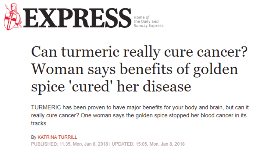
Kitchen cupboard remedies have become so mainstream that they become potentially dangerous when recommended for life-threatening diseases such as cancer. In fact, just a few months ago the Express asked “Can turmeric really cure cancer? Woman says benefits of golden spice ‘cured’ her disease”.

the express
Headline from the Express: “Can turmeric really cure cancer? Woman say benefits of golden spice ‘cured’ her disease”

But sometimes we wonder, if so many people believe it, maybe there’s really something in it?

In this series I will cover kitchen cupboard “cures” to investigate the claims made, and what the science really says. The series begins with the tasty Indian spice turmeric.
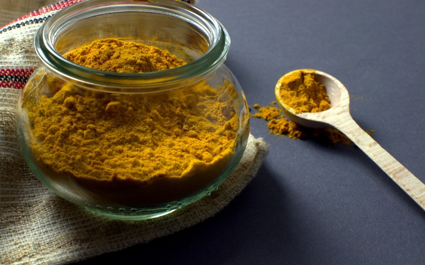
Turmeric

Turmeric is often reported as some sort of wonder spice. People who promote its use claim turmeric is anti-inflammatory, reduces cholesterol, treats diabetes, prevents Alzheimer’s disease and both prevents and cures cancer.

glass jar of orange coloured ground turmeric on a tea towel with a wooden spoon on the worktop next to it and turmeric on the spoon and counter

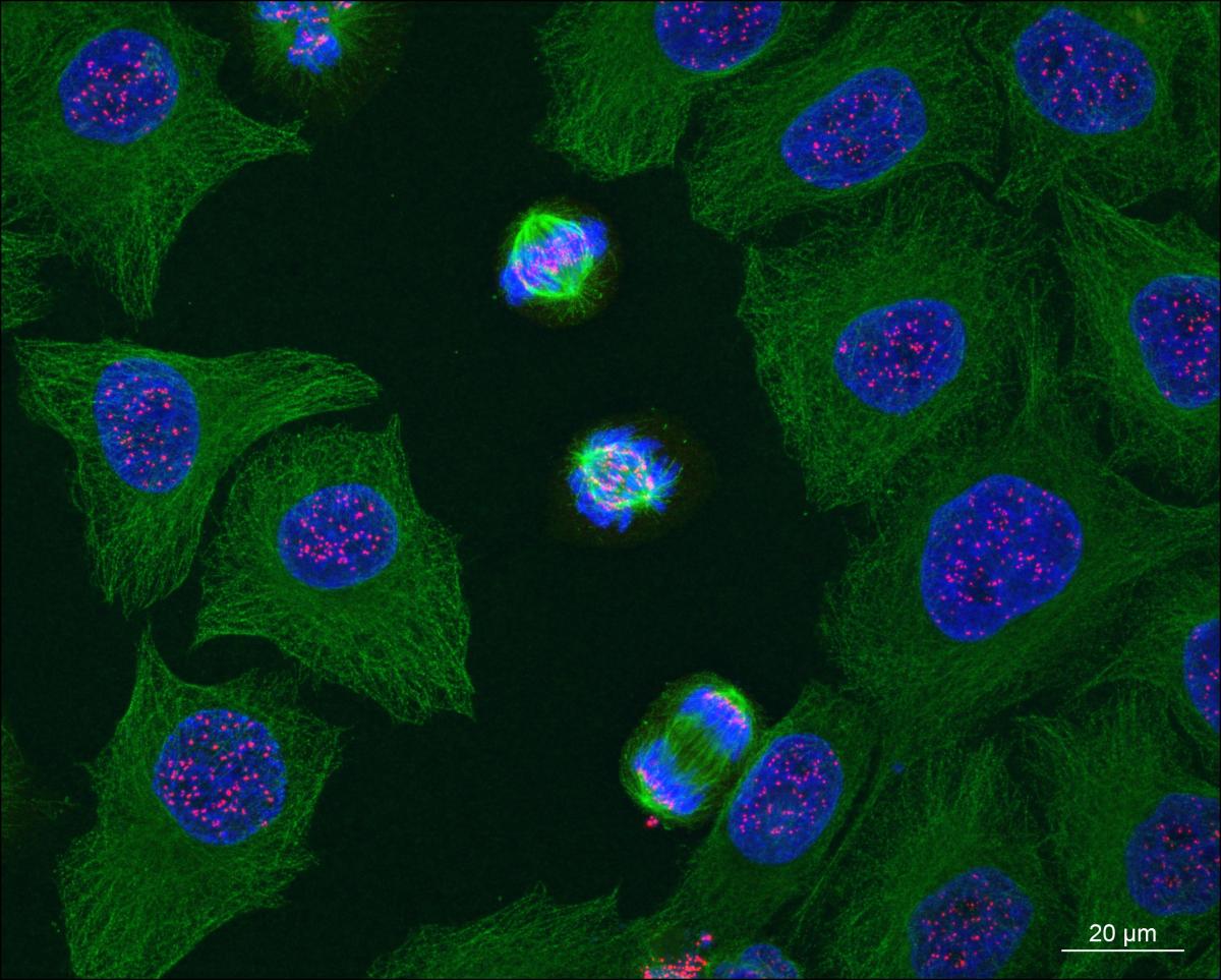
But actually, while this seems like a bizarre old wives’ tale, there is evidence supporting some of the claims for turmeric. The active ingredient in turmeric is curcumin or diferuloyl methane. Experiments in the lab show that curcumin alters the expression of genes in cells and some of these genes are related to specific pathways. For example, curcumin alters the expression of proteins related to inflammation in rat liver cells in a petri-dish and also alters the production of cholesterol in cells. Curcumin supplementation might also help manage some of the side effects of diabetes but only in conjunction with standard therapy. There is even some early evidence from mice with Alzheimer’s disease that curcumin can slow cognitive decline.

However, the science is also quite complicated. When it comes to cancer there is some evidence that curcumin can slow the growth of cancer cells in the lab but plenty of things slow cancer growth in the lab and never go on to prove useful therapies. Having said that, some clinical trials have shown that curcumin might one day prove useful as an adjunct to some cancer treatments in some cancer patients with some types of cancer.

The Express article above did discuss a case of a woman with myeloma, a type of blood cancer. This article was based on a single case published in British Medical Journal Reports in which a patient who had been on conventional treatment for many years suffered a relapse and was advised that there was nothing else doctors could do to help treat her cancer. The patient decided to take 8g chemical curcumin in tablet form per day in the hope it would treat her cancer. Her cancer has subsequently stabilised. This is a potentially interesting case – however it is only one single case that has been observed. Subsequent studies have not been done to investigate why this patient stabilised and there is insufficient evidence that it was the turmeric that was responsible. In fact, there are rare cases in which cancers such as myeloma can go into spontaneous remission without treatment and doctors believe this might be due to the patient’s own immune system targeting the cancer cells.

Important caveats

It would seem that the early research is fairly promising, however there are some very important caveats to remember here. So far, these studies are largely done in cells in a petri-dish or animals like mice or rats. We are not yet able to translate the findings to humans and we’re a long way from finding useful therapies using this compound.

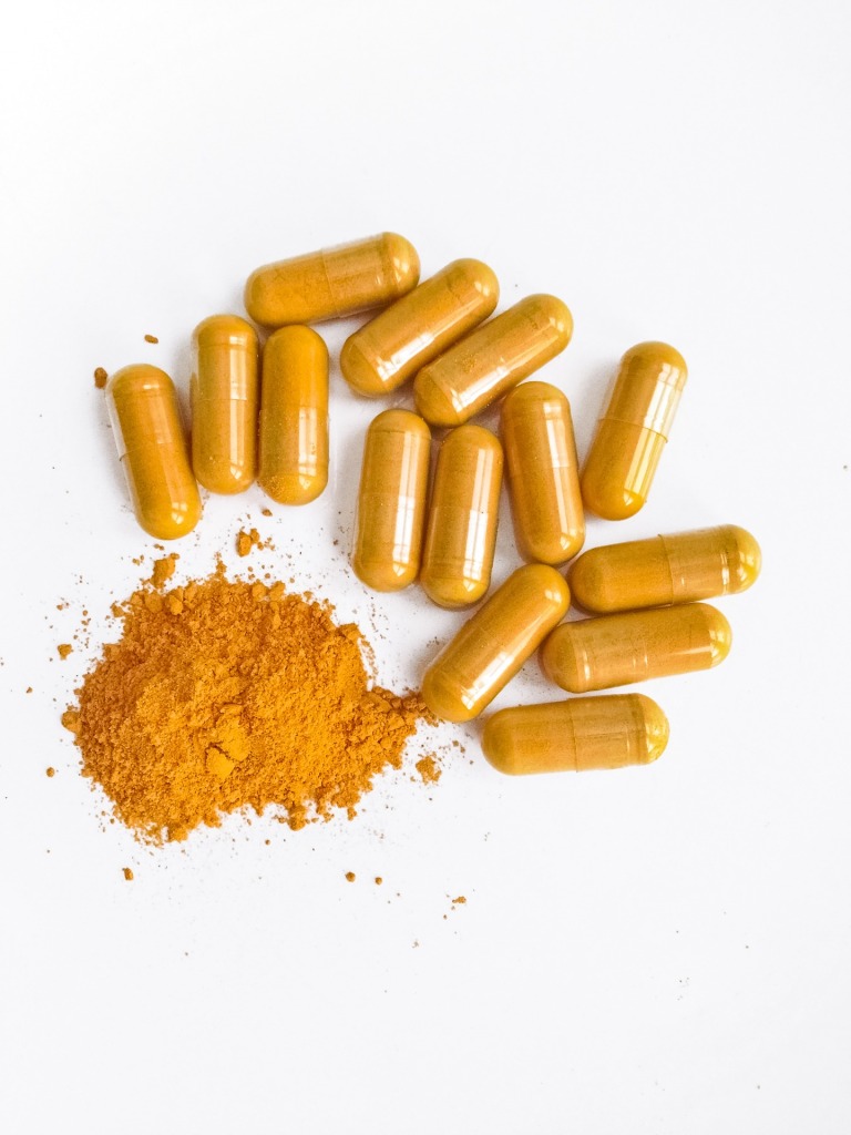
Importantly, the studies use chemical curcumin rather than dietary turmeric and usually have specific measured doses. If curcumin becomes a useful therapy, the dose will change between different diseases and different patients. There is no evidence supporting the use of turmeric in isolation to treat disease. In all studies it is used as a supplement to standard therapy.

Clear capsules with orange powder inside

Medical treatments should always be managed by a medical professional. Any ‘herbal’ remedy has the risk of interacting with conventional drugs. In the case of curcumin research has shown that the chemical can inhibit some cancer treatments so it is important we understand the role curcumin plays in reacting with other medications before using this to treat patients.

It is because of this risk of interaction with other medications that it is really important patients taking any herbal remedy supplements speak to their doctors about whether these supplements might harm themselves or the efficacy of their treatments. It is important to note that many supplements are not fully regulated and therefore may contain ingredients that cause harm. For example, some curcumin supplements have been shown to contain anti-inflammatory drugs which can cause liver damage if taken in excess.

Summary: while there is some early evidence the active ingredient of turmeric might one day prove a useful supplement to conventional therapy we’re a long way from this being clinically useful. We need much more research to confirm the efficacy of curcumin and to establish which compounds work best and at which doses.

Next week I’ll be writing about Rosemary. If you have any specific requests for a Kitchen Cupboard “Cure” for me to cover, please leave a comment or send me a tweet @AliceEmmaLouise.

For more information on turmeric and cancer you can see CRUK’s review.

Sources:


Cool science: using Zika virus to treat cancer?

Throughout the last two years there has been a great deal of news on the Zika virus – a virus spread by mosquitoes which was first identified in 1947 in Uganda. In a normal healthy adult Zika fever causes relatively mild symptoms or even none at all however the 2015-2016 Zika epidemic gave rise to widespread concern due to its propensity to cause microcephaly and brain defects in babies infected with the virus during development. The epidemic was declared over in late 2016 although there are still travel warnings to certain areas where the mosquitoes known to carry the virus are prevalent.

But research into this particular virus highlighted an interesting trait that we might be able to take advantage of – Zika has a preference for stem cells.

Zika and stem cells

The reason Zika virus is particularly dangerous in developing babies is that the virus causes damage in stem cells in the brain. A stem cell is a cell that isn’t yet programmed. They’re really important in development because when you create a baby you start with one sperm cell, one egg cell and these cells needs to combine, proliferate and then differentiate into all the different types of cell within the human body. Stem cells are unique cells that can be programmed or ‘differentiated’ into all sorts of different types of cells. Once a stem cell has differentiated it can’t turn back into a stem cell – a differentiated cell is committed to only ever being that cell type. The adult body has very, very few undifferentiated cells but a developing foetus has plenty. This explains the risk of Zika infection during pregnancy as Zika has been shown to target neural progenitor cells – a type of undifferentiated cell in the brain – that might lead to the microcephaly seen in babies infected with the virus during their development.

480px-Zika-chain-colored
Crystal structure of the Zika virus

Zika and brain cancer

Cancer stem cells are quite a complicated thing that I’m not going to try and do justice in this post because it’s a topic that deserves its own post. Scientists believe that some cancers do have associated cancer stem cells. How exactly cancer stem cells might contribute to cancer progression is far from fully understood. However, we do believe that the presence of cancer stem cells might contribute to cancer therapy relapse. This is particularly concerning in glioblastoma – an aggressive form of brain cancer, which has poor survival rates despite our best efforts. Without treatment, median survival is around 3 months from diagnosis. With treatment we are able to extend that survival to 12-15 months however the cancer usually recurs. Scientists believe this recurrence is all down to the presence of cancer stem cells.

The study

So here’s the clever part. Cancer researchers know we have a problem in treating glioblastoma. They also realised that Zika virus is a relatively mild virus, which attacks stem cells. The adult brain doesn’t really have stem cells – unless the adult has glioblastoma. Cancer stem cells in the brain lead to cancer relapse; Zika attacks brain stem cells. Maybe we can make use of these two pieces of information.

So, scientists did some experiments. Firstly, they took some glioblastoma cells from patient tumours and they grew them in a dish in the lab. Then they infected them with Zika virus. They looked at either glioblastoma differentiated cells or glioblastoma stem cells. And they looked at the infection rate. Over 48 hours, over 60% of the stem cells were infected and this increased over time as the virus spread. The differentiated cells were infected too but not as much. What was especially interesting was that the stem cells infected with the virus had severely reduced ability to multiply and they had an increase in cell death. This was specific to only the stem cells and didn’t affect the differentiated cells. The virus kills cancer stem cells and prevents them from spreading.

Next the scientists took some patient tissue samples – this allowed them to look at a whole mixture of cells in a slightly more normal context without having to infect patients. They infected the tissue samples with Zika and saw that cancer samples were infected successfully and the virus only hit the stem cells and not the other cell types in the sample. They also looked at some brain samples from epilepsy patients and the virus didn’t infect them showing that the virus really is specific for stem cells!

jem.20171093
Glioblastoma tissue sample (from the paper)


Finally, they used the virus to treat mouse models of glioblastoma. They took mice with glioblastoma tumours in the brain and infected them with the virus. They saw that the tumours were much smaller and the mouse had improved survival when they were infected with the virus compared to control treated mice. They went on to show that they got an even better effect when combined with other glioblastoma treatments.

Benefits

Current treatments have two problems when it comes to glioblastoma. Firstly, the cancer stem cells make recurrence almost inevitable. This could drastically improve average survival times. Secondly, all brain cancer treatments have to be able to cross the blood-brain barrier in order to get through to the cancer cells. The blood brain barrier is an important way to keep things out of the brain where they might cause damage but it also serves as a way to keep brain tumours trapped in and harder to treat. Zika is great at crossing the blood-brain barrier.

What next?

We’re still such a long way from this being a useful patient treatment. In order to use this as a treatment we need to modify the virus in such a way that it will not spread from person to person and it will not cause the patient any harm. Currently virus work is always done in very specialised laboratories with expert training on how to prevent spread and with many, many precautions. If it were to be used as a therapy we’d need lots and lots of precautions to make sure it were safe. So far this has only been done in lab grown cells (albeit ones taken from patients mouse models of cancer. But it’s incredibly interesting research and a great example of how cancer research is so quick to develop and understand how we can take advantage of what we know about the disease and use that to treat it.


Please let me know in the comments if you’d like to see a post on any of the topics from this post – Zika virus, glioblastoma, stem cells?

If you found this interesting, please share it with three other people who might find it interesting too! Sharing cool cancer research gives us all a little more hope!


Image credit for the crystal structure of Zika: By Manuel Almagro Rivas – Own work, CC BY-SA 4.0, https://commons.wikimedia.org/w/index.php?curid=47941048

Cool Science: taking advantage of cancer cell biology for cancer treatment

One of the big problems when it comes to treating cancer using drugs is that these drugs flood the patient’s body and cause detrimental side effects when they reach areas other than the malignant tumour.  Cancer cells are derived from our own healthy cells – they’re hard to target specifically without also hitting our healthy cells. A lot of research goes into trying to get around this problem. I came across a particularly interesting study that I thought I’d share here.

Earlier this year Sofie Snipstad et al. published a paper in Ultrasound in Medicine & Biology titled “Ultrasound improves the delivery and therapeutic effect of nanoparticle-stabilized microbubbles in breast cancer xenografts”. This paper was particularly cool because of the rationale behind it. Nanoparticle delivery of drugs is something scientists have been working on for a little while. The premise is that you take your drug and you wrap it up inside a small protective bubble allowing the drug to travel to a specific site before it is released. Due to a quirk of cancer biology, this is particularly great as a cancer therapy. When tumours grow, they start to form their very own blood supply – only the blood vessels that they grow are more leaky than normal blood vessels. They allow slightly larger molecules to pass through from the blood vessel and into the surrounding tumour.  This means we can use nanoparticles to deliver cancer drugs specifically into tumour sites by allowing the particles to travel through these leaky blood vessels. But then we hit another problem – if the tumour blood supply doesn’t reach the very depths of the tumour then the particles are too big to get all the way through. You can treat the edges but not the very centre of the tumour. So, this paper worked on a special combination – they took a bunch of nanoparticles containing chemotherapy and they bundled them up together into microbubbles that can travel around the blood system easily and safely until they reach the tumour site. Then, the researchers used a focused shot of ultrasound to break up the bubbles and release the nanoparticles. This also served to allow gentle tissue massage by the ultrasound to allow the nanoparticles to distribute further throughout the tumour. Once in the tumour, the cancer cells start to take up the nanoparticles and inside the cell the drug is released and can kill the cancer cell from the inside.

Picture1Image: figure from the paper

Any microbubbles that weren’t in the tumour site and therefore not exposed to the focused ultrasound could be easily cleared from the body without releasing the drug which means the only cells targeted by the therapy are the cancer cells. This means we can hit the cancer cells with a higher, more toxic dose because the healthy cells are not going to be hit with the same dose.

The researchers in this paper were testing the optimal way of doing this in mice suffering with triple negative breast cancer – one of the more aggressive forms of the disease – with positive results. The mouse tumours took up the drug 2.3x better and there was no tissue damage identified. All of the tumours either regressed or else the mice went into complete remission. The authors described this as a “promising proof-of-concept study”.

New Study: Does chemotherapy promote cancer metastasis?

A little while ago my attention was drawn to an article published in July 2017 on a blog called The Mind Unleashed titled “Chemotherapy to Spread Cancer, Cause Lethal Tumours in Groundbreaking New Study”. The article reported on a paper published the same month in the journal of Science Translational Medicine. This article claimed that the researchers had “proven that chemotherapy causes cancer cells to spread throughout the body – to replicate themselves, making your cancer worse, not better”.

This is a frightening thought. Could it be possible that in treating cancer we are actually promoting its survival? As a cancer researcher I was very, very sceptical of this claim. Chemotherapy is a cancer treatment that has been available for a long time. It was first studied in the early 20th century and first administered in 1942. On the day of writing there are over 2.9 million papers referencing the term chemotherapy in the title, key words or abstract available on PubMed (a database for published, peer-reviewed material). We have done an incredible amount of research into different types of chemotherapy treatments and those that work are the only ones approved for use in the clinic.

That’s not to say we know everything about chemotherapy, though. The human body is a complicated thing, as is cancer. We might well be missing something. The most important part of research is to continually progress, to follow the science where it leads us in the most unbiased way possible.

*

The paper in question is one reporting a study undertaken by researchers at Albert Einstein College of Medicine in the US. The authors were working on a particular chemotherapeutic agent called paclitaxel (brand name Taxol) in mouse models of breast cancer.

Paclitaxel is a drug derived, originally, from the bark of the Pacific Yew tree. It was first discovered by a screen on plant derivatives undertaken by the National Cancer Institute in the early 1960’s. After decades of research into the chemical – first isolating the chemical structure and later the mechanism of action – paclitaxel was approved by the FDA in 1992. Today, we make it from a precursor taken from the needles of this plant. We are unable to produce it entirely synthetically.

Paclitaxel works – it prolongs progression free survival and shrinks tumours. The mechanism of action is well established; paclitaxel binds to a protein involved in cell division thus preventing tumour cell replication. Consequently those cancer cells die and tumour size is reduced.

cancer cells, cell division, cancer research

There are, of course, side effects to any drug and paclitaxel does have a number of them. In particular, paclitaxel is not soluble in water so for treatment it must be diluted in a derivative of castor oil. This is not particularly well tolerated by the human body and patients must be co-treated with corticoids and antihistamines to prevent dangerous hypersensitivity reactions so research is ongoing into better ways of administering paclitaxel.

One avenue of research has been into a particular quirk of paclitaxel treatment. It has been identified by clinical trial that when paclitaxel is used prior to primary treatment (such as surgery) in breast cancer (known as a neoadjuvent therapy) the survival rate of patients is not increased beyond that of surgery alone. This is despite a reduction in tumour size when using paclitaxel as a neoadjuvent. If tumour size is reduced you might expect survival to be enhanced – there was a discrepancy here not currently explained by the available science.

The authors of this paper had a theory. They knew that paclitaxel treatment is associated with an increase in a particular type of immune cell called macrophages moving into the tumour site. They also knew that macrophages might be involved in metastasis. Therefore, they posited that perhaps the reason paclitaxel didn’t prolong survival despite shrinking the tumour might be because the tumour was able to sustain itself by travelling elsewhere in the body.

They investigated this question in a number of laboratory mice which suffer from breast cancer and are used as models of the disease in humans. The researchers found that there was an increase in markers for metastasis in those breast cancer mice treated with paclitaxel and an increase in circulating cancer cells in the bloodstream. And then they took it another step forward. The authors noticed that the increase in metastasis markers included an increase in a protein called TIE2. They had reason to believe that this was an important part of the problem so they co-treated the mice with an inhibitor against TIE2. This they showed reduced the markers of metastasis and the circulating cancer cells in the blood.

mouse model, science, laboratory, cancer research

The important conclusion of this study was not that paclitaxel might promote metastasis in mice with breast cancer. It was that this particular type of chemotherapy might have another negative effect we didn’t know about. It is important to know about this because now we can monitor patients for changes in their metastasis markers when they are treated with this type of chemotherapy and we can switch them on to a different form of treatment if necessary – or we can co-treat them with TIE2 inhibitors. The most important thing in medicine is to have as much knowledge as we possibly can. We shouldn’t be fearful of negative effects of good treatments – unfortunately to kill cancer cells in the body will have some negative effects. But we need to be aware of them and we need to manage them carefully and make sure we put the needs of patients first. Research like this helps us make informed decisions on treatment options. Of course it requires further research first in more animal models and later in humans but it a stepping stone to giving us important information that may well help us to save more lives.

*

What worries me more is that blogs like The Unleashed Mind misrepresent the data reported and promote distrust in reliable medical research and the scientific method in general. Anyone communicating the findings of academic research has a responsibility to represent it accurately especially when that communication might well influence a patient’s decision when it comes to their health. A cornerstone of medicine is to give patients the opportunity to informed consent – we should all endeavour to present the information as accurately as is possible.

photo credit: ZEISS Microscopy