Will we ever cure HIV infection?

So far on this blog, I’ve talked a lot about cancer. I’ve been working in cancer cell biology for a number of years, and it’s an area that a lot of people are interested in, so it makes for a sensible topic. By training, though, I am a cellular and molecular physiologist. I’m interested in how all cells work; they don’t have to be cancer cells.

The human body, and cells in particular, amaze me. I love learning the intricate details of exactly how our cells work on a molecular level, but I love how we can identify little changes that might be useful in understanding disease and treatment even more.

Earlier this year I came across an interesting study on treating individuals infected with the Human Immunodeficiency Virus (HIV). HIV is a complicated virus and has long been a source of fear. But by understanding the science of the virus we can look to treat infection and perhaps dispel some of those fears. So here’s a bit about HIV, and then a bit about a cool study.

HIV

HIV is a type of virus that we call a retrovirus. A retrovirus carries its genetic material as a single strand of RNA, instead of the double-strand of DNA we use as humans. To transmit its genetic material the virus needs to construct DNA, which it does inside a host cell. In the case of HIV, the host cells are cells within the human immune system – white blood cells called helper T cells.  Once an HIV molecule (a virion) gets inside a human host cell, it uses an enzyme within the virus to transcribe a piece of its RNA into DNA. It then slots that DNA into the host cell genome. It can remain dormant for many years but eventually the host cell machinery produces many, many copies of this piece of DNA, releasing more HIV virions into the blood stream. Essentially, the virus hijacks the host cell’s normal processes in order to make copies of itself.

Picture1
The life cycle of HIV: the virus enters a host cell and releases its RNA which is transcribed into viral DNA. Viral DNA is integrated into the host cell DNA where it can lie dormant for many years. Upon activation, the host cell produces viral RNA allowing the generation of new virions which are released into the blood and are able to infect new host cells.  Image taken from Encyclopaedia Britannica.

The HIV virus does not need all the machinery required for its own replication therefore the HIV genome is tiny, consisting of only nine genes. By comparison, the human genome consists of around 20,000 genes!

Treating HIV

These days we are pretty good at treating and managing HIV infection. HIV infection isn’t currently curable, but we’re so good at treating it that many patients live a long, healthy life while carrying HIV. There are many caveats to that, including political problems related to getting treatment to all the parts of the world that need it.

The very first treatment we produced for HIV was an inhibitor that blocked the action of a protein important in converting the virus’ RNA into DNA, so preventing its subsequent replication and spread throughout the body. Since then we’ve designed different types of antiretroviral therapy (ART) which target different processes within the virus’ lifecycle. We can stop the virus getting into a cell, stop its RNA converting to DNA, stop that DNA integrating into the human genome and stop the virus from assembling and breaking out of the host cell. The problem is, once the virus is integrated into the host cell DNA it can lay dormant, untouched by inhibitors even when the viral load is reduced in the patients’ blood. We have to maintain treatment to prevent this dormant DNA from activating and the virus spreading around the human body. It is possible, too, for the virus become resistant to each therapy meaning careful assessment of the patient and modification of the therapy accordingly. In this way, we can manage the infection long-term. But what if we could figure out a way to target the dormant virus sitting in host cell DNA?

New study

Picture2
Title and authors of the new study published in September 2017 in PLOS Pathogens

A new study published in September aimed to do exactly this. Earlier research had shown that a particular type of drug can ‘activate’ the host-cells which hold dormant HIV DNA. These ‘activated’ cells produce lots and lots of RNA for the virus which “kicks” the hiding virus back into action. Using this principle, the standard anti-retroviral drugs are then able to “kill” the virus specifically targeting this pool of dormant virus that standard therapy alone is unable to target. This strategy is referred to as “kick and kill”. While other researchers have studied this principle before, the effect on host-cell activation has been minimal which means only a small proportion of cells holding dormant HIV can be targeted. The larger this proportion, the closer we can get to activating and killing every cell holding dormant-HIV and the closer we can get to curing HIV infection.

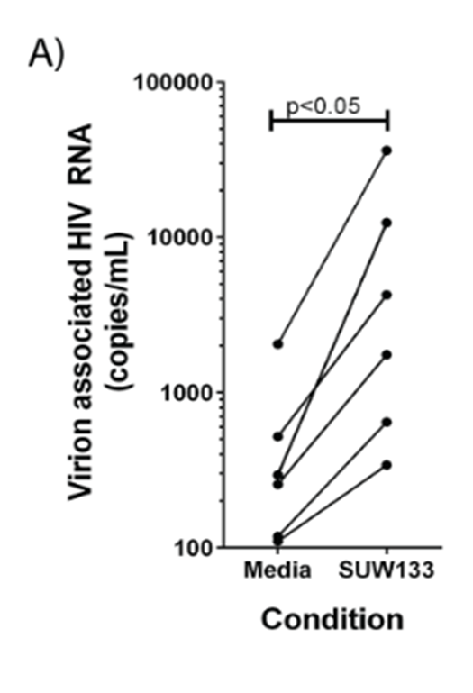
In this study the authors used a synthetic compound, SUW133, which would they hoped might activate more host cells and therefore be more successful.  Their initial work looked at the effectiveness of the compound in cells taken from six patients. They showed that control treated cells (treated with only media) had relatively few RNA molecules from the HIV virus whereas when they took those same patient-derived cells and treated them with the SUW133 the number of viral RNA molecules increased significantly. This indicates that SUW133 does, indeed, activate the dormant virus and kicks the host cells into action.

Picture3
Data taken from the paper. Comparison of control treated human patient cells with SUW133 treated human patient cells. Samples derived from the same individual patients are connected with a line showing the increase in activation of cells per patient when treated with SUW133.

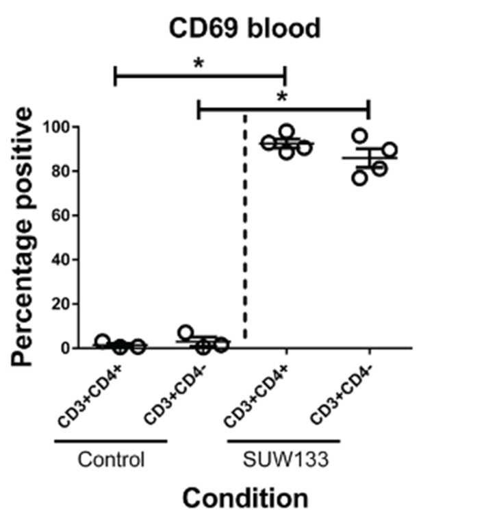
They then wanted to look at what would happen in a living organism, so the researchers had to infect humanised mice (mice genetically modified to express human genes) with HIV. They did this for 3-4 weeks, after which they treated the mice with antiretroviral therapy for 2-5 weeks. Once they were sure that only dormant HIV would remain in the mice, they treated them with their special synthetic compound and looked in the blood for evidence of HIV-host-cell activation. In mice treated with a control (inert) compound they found relatively low proportions of specific cell types (CD3+/CD4+ and CD3+/CD4-) that expressed a marker for activation (CD69) however upon treatment with the active compound (SUW133) the proportion of “activated” CD69 positive cells rose dramatically.

Picture4
Data from the paper. The proportion of  activated (CD69+) blood cells in the blood of HIV positive mice is much higher in mice treated with SUW133 compared to control treated mice.

What’s more, the researchers showed that a small proportion (around 25%) of these activated cells were dead or dying which shows promise for the additional treatment with standard antiretroviral therapy to fully “kill” the virus.

Overall the authors have shown that they can activate dormant HIV which they hope will give us the opportunity to kill this currently untreatable pool of the virus and one day give us a cure to HIV infection.

What’s clear is that this is still early days – the researchers need to fully establish the effect of this “kick” strategy when combined with the “kill” treatment (ART). However, it is incredibly promising that we might someday be able to activate host-cells carrying dormant HIV genetic material so that we can remove those cells entirely. If we are successful, we could go from long-term HIV infection management to HIV-infection cure.


Science or Pseudoscience: Ancient Chinese Calligraphy Ink and Cancer Treatment

When it comes to reading about cancer cures in the media it is usually good advice to be pretty skeptical. Sometimes the story lends well to skepticism – like this one recently covered in a few outlets where it was proclaimed that scientists had found a cure for cancer in ancient Chinese calligraphy ink. It’s a story that has everything – including lasers. Here is a romantic idea that the cure for cancer might lie in an ink that is produced from plants and has been handed down between generations as a way to share writing and art. The proposed treatment is billed as non-invasive and specific only for cancer cells – could this really be science?

I was particularly suspicious when I saw that the representation of the story on Natural News, a prominent proponent of pseudoscience, looked a lot like the story on Science Alert which is typically more of a reliable source on science stories. So I did some digging into the data for this finding.

The paper that was covered in the media is titled “New Application of Old Material: Chinese Traditional Ink for Photothermal Therapy of Metastatic Lymph Nodes” and was published in the journal ACS Omega in August 2017. The researchers were based in Shanghai distributed across various institutes at different hospitals or universities. They had been working on a relatively new cancer therapy called Photo-thermal Therapy (PTT).

Photo-thermal therapy (PTT)

In cancer treatment, PTT is the use of specific types of nanoparticles to generate cell damaging heat specifically at the tumour sites. Scientists take a material that can be stimulated with light – typically infra-red – causing the generation of heat and subsequent tumour cell death. The problem is that many of these nanoparticles are toxic or expensive so in order to make the treatment as efficient and effective as possible, we need to find just the right material.

Hu-Kaiwen

Hu-Kaiwen (Hu-ink) is an ink that has been used by calligraphers in China for hundreds of years. It’s derived from plants, is mostly made up of carbon and it is black in colour which the materials used for PTT tend to be. So scientists got to wondering if it might be useful for PTT.

Firstly the authors of this study looked at the stability of the ink. They diluted it in different things including water and saline and made sure it was stable when stored over time. They looked at the structure of the ink and noticed that it typically forms small aggregates of 20-50nm in diameter – nanoparticles. They confirmed that the core component of the ink was carbon and they stimulated different concentrations of the diluted ink with infra-red lasers and tested the temperature. They found that Hu-ink was more efficient at converting light into heat than most other PTT materials reaching temperatures of 55°C after five minutes irradiation.

Hu-ink for PTT in cancer cells grown in the lab

In research we use cell models of cancer which we refer to as cancer cell lines. These are cells that have originally been taken from patients with different types of cancer but are grown and stored artificially in the lab. We use them so we can test things out on cells that behave like cancer but aren’t in a human body before we move on to doing tests in living organisms. The researchers in this study used some cancer cells originally derived from colon (SW-620) and colorectal (HCT-116) cancer. First they treated the cells with just the Hu-ink and the cells tolerated it really well proving that the ink solution itself wasn’t toxic. Then they treated the cells with the ink and combined it with irradiation with the infra-red laser.

Hu-ink
HCT-116 cells treated with Hu-ink and infra-red compared to HCT-116 cells treated with infra-red only. Image modified from the paper.

The image above is part of a figure taken from the paper itself. There are three images of HCT-116 colorectal cancer cells. In the top image, the cells were treated with only the laser for five minutes. They were not given any Hu-ink. In the middle image the cells were treated both with the Hu-ink at a medium dose for two hours and the laser for five minutes. In the bottom image the cells received a high dose of the Hu-ink for two hours and the laser for five minutes. The red cells are cells stained with a marker of cell death. The green cells are living cells. You can also see that the dead cells are much smaller than the living cells. In the top image where cells did not receive any ink – all of the cells are alive. In the bottom image where the cells had a high dose of ink – all of the cells are dead. In the middle image with a lower dose of the ink, there are some living cells and some dead cells. In other words – when you treat cancer cells with both Hu-ink and infra-red, the cancer cells die.

Hu-ink for PTT in mice

The researchers wanted to be sure that this technique was safe in living organisms and that it was able to kill cancer cells in living organisms. So they took mice with cancer of the lymph nodes and injected Hu-ink into the tumour which they then irradiated with infra-red. After an allocated treatment time, they removed the tumours from the mice and measured them.

Hu-ink2
Lymph node tumours taken from mice treated with Hu-ink and infra-red versus control treatments. Image modified from the paper.

For this experiment they had three control conditions – NS (the tumours were injected with saline), NS plus laser (the tumours were injected with saline and treated with a laser) and Hu-ink (the cells were injected with Hu-ink). They had one test condition – the Hu-ink plus laser. The test condition is the only one that the authors predicted would have an effect on tumour size. And that’s exactly what they saw. Lymph node tumours treated with Hu-ink and infra-red were significantly smaller than the control conditions. They also saw that the surrounding tissue wasn’t damaged suggesting that the treatment is safe.

Study conclusion

This study is a proof-of-principle study. The authors have shown that this ink can be used in PTT therapy with a positive effect and in a safe way in mice and in the lab. It is a very small scale study and it is a single study. It needs replicating before we can be confident in the result and it needs to be studied on a larger scale and have many more safety tests before we could begin to think about using it in patients. But it is a really promising study. It takes a treatment we already use and aims to make that treatment safer and cheaper and more available to patients. It is a non-invasive treatment and it can be used in ways that reduces tissue damage to healthy tissue while targeting cancer cells for cell death. It’s a great example of some really cool science, using materials that have been available for many years and applying them to modern techniques.

It might seem far-fetched to say ancient ink can help us treat cancer, but really it’s just cool science!

Cool science: using Zika virus to treat cancer?

Throughout the last two years there has been a great deal of news on the Zika virus – a virus spread by mosquitoes which was first identified in 1947 in Uganda. In a normal healthy adult Zika fever causes relatively mild symptoms or even none at all however the 2015-2016 Zika epidemic gave rise to widespread concern due to its propensity to cause microcephaly and brain defects in babies infected with the virus during development. The epidemic was declared over in late 2016 although there are still travel warnings to certain areas where the mosquitoes known to carry the virus are prevalent.

But research into this particular virus highlighted an interesting trait that we might be able to take advantage of – Zika has a preference for stem cells.

Zika and stem cells

The reason Zika virus is particularly dangerous in developing babies is that the virus causes damage in stem cells in the brain. A stem cell is a cell that isn’t yet programmed. They’re really important in development because when you create a baby you start with one sperm cell, one egg cell and these cells needs to combine, proliferate and then differentiate into all the different types of cell within the human body. Stem cells are unique cells that can be programmed or ‘differentiated’ into all sorts of different types of cells. Once a stem cell has differentiated it can’t turn back into a stem cell – a differentiated cell is committed to only ever being that cell type. The adult body has very, very few undifferentiated cells but a developing foetus has plenty. This explains the risk of Zika infection during pregnancy as Zika has been shown to target neural progenitor cells – a type of undifferentiated cell in the brain – that might lead to the microcephaly seen in babies infected with the virus during their development.

480px-Zika-chain-colored
Crystal structure of the Zika virus

Zika and brain cancer

Cancer stem cells are quite a complicated thing that I’m not going to try and do justice in this post because it’s a topic that deserves its own post. Scientists believe that some cancers do have associated cancer stem cells. How exactly cancer stem cells might contribute to cancer progression is far from fully understood. However, we do believe that the presence of cancer stem cells might contribute to cancer therapy relapse. This is particularly concerning in glioblastoma – an aggressive form of brain cancer, which has poor survival rates despite our best efforts. Without treatment, median survival is around 3 months from diagnosis. With treatment we are able to extend that survival to 12-15 months however the cancer usually recurs. Scientists believe this recurrence is all down to the presence of cancer stem cells.

The study

So here’s the clever part. Cancer researchers know we have a problem in treating glioblastoma. They also realised that Zika virus is a relatively mild virus, which attacks stem cells. The adult brain doesn’t really have stem cells – unless the adult has glioblastoma. Cancer stem cells in the brain lead to cancer relapse; Zika attacks brain stem cells. Maybe we can make use of these two pieces of information.

So, scientists did some experiments. Firstly, they took some glioblastoma cells from patient tumours and they grew them in a dish in the lab. Then they infected them with Zika virus. They looked at either glioblastoma differentiated cells or glioblastoma stem cells. And they looked at the infection rate. Over 48 hours, over 60% of the stem cells were infected and this increased over time as the virus spread. The differentiated cells were infected too but not as much. What was especially interesting was that the stem cells infected with the virus had severely reduced ability to multiply and they had an increase in cell death. This was specific to only the stem cells and didn’t affect the differentiated cells. The virus kills cancer stem cells and prevents them from spreading.

Next the scientists took some patient tissue samples – this allowed them to look at a whole mixture of cells in a slightly more normal context without having to infect patients. They infected the tissue samples with Zika and saw that cancer samples were infected successfully and the virus only hit the stem cells and not the other cell types in the sample. They also looked at some brain samples from epilepsy patients and the virus didn’t infect them showing that the virus really is specific for stem cells!

jem.20171093
Glioblastoma tissue sample (from the paper)


Finally, they used the virus to treat mouse models of glioblastoma. They took mice with glioblastoma tumours in the brain and infected them with the virus. They saw that the tumours were much smaller and the mouse had improved survival when they were infected with the virus compared to control treated mice. They went on to show that they got an even better effect when combined with other glioblastoma treatments.

Benefits

Current treatments have two problems when it comes to glioblastoma. Firstly, the cancer stem cells make recurrence almost inevitable. This could drastically improve average survival times. Secondly, all brain cancer treatments have to be able to cross the blood-brain barrier in order to get through to the cancer cells. The blood brain barrier is an important way to keep things out of the brain where they might cause damage but it also serves as a way to keep brain tumours trapped in and harder to treat. Zika is great at crossing the blood-brain barrier.

What next?

We’re still such a long way from this being a useful patient treatment. In order to use this as a treatment we need to modify the virus in such a way that it will not spread from person to person and it will not cause the patient any harm. Currently virus work is always done in very specialised laboratories with expert training on how to prevent spread and with many, many precautions. If it were to be used as a therapy we’d need lots and lots of precautions to make sure it were safe. So far this has only been done in lab grown cells (albeit ones taken from patients mouse models of cancer. But it’s incredibly interesting research and a great example of how cancer research is so quick to develop and understand how we can take advantage of what we know about the disease and use that to treat it.


Please let me know in the comments if you’d like to see a post on any of the topics from this post – Zika virus, glioblastoma, stem cells?

If you found this interesting, please share it with three other people who might find it interesting too! Sharing cool cancer research gives us all a little more hope!


Image credit for the crystal structure of Zika: By Manuel Almagro Rivas – Own work, CC BY-SA 4.0, https://commons.wikimedia.org/w/index.php?curid=47941048

Cool Science: taking advantage of cancer cell biology for cancer treatment

One of the big problems when it comes to treating cancer using drugs is that these drugs flood the patient’s body and cause detrimental side effects when they reach areas other than the malignant tumour.  Cancer cells are derived from our own healthy cells – they’re hard to target specifically without also hitting our healthy cells. A lot of research goes into trying to get around this problem. I came across a particularly interesting study that I thought I’d share here.

Earlier this year Sofie Snipstad et al. published a paper in Ultrasound in Medicine & Biology titled “Ultrasound improves the delivery and therapeutic effect of nanoparticle-stabilized microbubbles in breast cancer xenografts”. This paper was particularly cool because of the rationale behind it. Nanoparticle delivery of drugs is something scientists have been working on for a little while. The premise is that you take your drug and you wrap it up inside a small protective bubble allowing the drug to travel to a specific site before it is released. Due to a quirk of cancer biology, this is particularly great as a cancer therapy. When tumours grow, they start to form their very own blood supply – only the blood vessels that they grow are more leaky than normal blood vessels. They allow slightly larger molecules to pass through from the blood vessel and into the surrounding tumour.  This means we can use nanoparticles to deliver cancer drugs specifically into tumour sites by allowing the particles to travel through these leaky blood vessels. But then we hit another problem – if the tumour blood supply doesn’t reach the very depths of the tumour then the particles are too big to get all the way through. You can treat the edges but not the very centre of the tumour. So, this paper worked on a special combination – they took a bunch of nanoparticles containing chemotherapy and they bundled them up together into microbubbles that can travel around the blood system easily and safely until they reach the tumour site. Then, the researchers used a focused shot of ultrasound to break up the bubbles and release the nanoparticles. This also served to allow gentle tissue massage by the ultrasound to allow the nanoparticles to distribute further throughout the tumour. Once in the tumour, the cancer cells start to take up the nanoparticles and inside the cell the drug is released and can kill the cancer cell from the inside.

Picture1Image: figure from the paper

Any microbubbles that weren’t in the tumour site and therefore not exposed to the focused ultrasound could be easily cleared from the body without releasing the drug which means the only cells targeted by the therapy are the cancer cells. This means we can hit the cancer cells with a higher, more toxic dose because the healthy cells are not going to be hit with the same dose.

The researchers in this paper were testing the optimal way of doing this in mice suffering with triple negative breast cancer – one of the more aggressive forms of the disease – with positive results. The mouse tumours took up the drug 2.3x better and there was no tissue damage identified. All of the tumours either regressed or else the mice went into complete remission. The authors described this as a “promising proof-of-concept study”.

New Study: Does chemotherapy promote cancer metastasis?

A little while ago my attention was drawn to an article published in July 2017 on a blog called The Mind Unleashed titled “Chemotherapy to Spread Cancer, Cause Lethal Tumours in Groundbreaking New Study”. The article reported on a paper published the same month in the journal of Science Translational Medicine. This article claimed that the researchers had “proven that chemotherapy causes cancer cells to spread throughout the body – to replicate themselves, making your cancer worse, not better”.

This is a frightening thought. Could it be possible that in treating cancer we are actually promoting its survival? As a cancer researcher I was very, very sceptical of this claim. Chemotherapy is a cancer treatment that has been available for a long time. It was first studied in the early 20th century and first administered in 1942. On the day of writing there are over 2.9 million papers referencing the term chemotherapy in the title, key words or abstract available on PubMed (a database for published, peer-reviewed material). We have done an incredible amount of research into different types of chemotherapy treatments and those that work are the only ones approved for use in the clinic.

That’s not to say we know everything about chemotherapy, though. The human body is a complicated thing, as is cancer. We might well be missing something. The most important part of research is to continually progress, to follow the science where it leads us in the most unbiased way possible.

*

The paper in question is one reporting a study undertaken by researchers at Albert Einstein College of Medicine in the US. The authors were working on a particular chemotherapeutic agent called paclitaxel (brand name Taxol) in mouse models of breast cancer.

Paclitaxel is a drug derived, originally, from the bark of the Pacific Yew tree. It was first discovered by a screen on plant derivatives undertaken by the National Cancer Institute in the early 1960’s. After decades of research into the chemical – first isolating the chemical structure and later the mechanism of action – paclitaxel was approved by the FDA in 1992. Today, we make it from a precursor taken from the needles of this plant. We are unable to produce it entirely synthetically.

Paclitaxel works – it prolongs progression free survival and shrinks tumours. The mechanism of action is well established; paclitaxel binds to a protein involved in cell division thus preventing tumour cell replication. Consequently those cancer cells die and tumour size is reduced.

cancer cells, cell division, cancer research

There are, of course, side effects to any drug and paclitaxel does have a number of them. In particular, paclitaxel is not soluble in water so for treatment it must be diluted in a derivative of castor oil. This is not particularly well tolerated by the human body and patients must be co-treated with corticoids and antihistamines to prevent dangerous hypersensitivity reactions so research is ongoing into better ways of administering paclitaxel.

One avenue of research has been into a particular quirk of paclitaxel treatment. It has been identified by clinical trial that when paclitaxel is used prior to primary treatment (such as surgery) in breast cancer (known as a neoadjuvent therapy) the survival rate of patients is not increased beyond that of surgery alone. This is despite a reduction in tumour size when using paclitaxel as a neoadjuvent. If tumour size is reduced you might expect survival to be enhanced – there was a discrepancy here not currently explained by the available science.

The authors of this paper had a theory. They knew that paclitaxel treatment is associated with an increase in a particular type of immune cell called macrophages moving into the tumour site. They also knew that macrophages might be involved in metastasis. Therefore, they posited that perhaps the reason paclitaxel didn’t prolong survival despite shrinking the tumour might be because the tumour was able to sustain itself by travelling elsewhere in the body.

They investigated this question in a number of laboratory mice which suffer from breast cancer and are used as models of the disease in humans. The researchers found that there was an increase in markers for metastasis in those breast cancer mice treated with paclitaxel and an increase in circulating cancer cells in the bloodstream. And then they took it another step forward. The authors noticed that the increase in metastasis markers included an increase in a protein called TIE2. They had reason to believe that this was an important part of the problem so they co-treated the mice with an inhibitor against TIE2. This they showed reduced the markers of metastasis and the circulating cancer cells in the blood.

mouse model, science, laboratory, cancer research

The important conclusion of this study was not that paclitaxel might promote metastasis in mice with breast cancer. It was that this particular type of chemotherapy might have another negative effect we didn’t know about. It is important to know about this because now we can monitor patients for changes in their metastasis markers when they are treated with this type of chemotherapy and we can switch them on to a different form of treatment if necessary – or we can co-treat them with TIE2 inhibitors. The most important thing in medicine is to have as much knowledge as we possibly can. We shouldn’t be fearful of negative effects of good treatments – unfortunately to kill cancer cells in the body will have some negative effects. But we need to be aware of them and we need to manage them carefully and make sure we put the needs of patients first. Research like this helps us make informed decisions on treatment options. Of course it requires further research first in more animal models and later in humans but it a stepping stone to giving us important information that may well help us to save more lives.

*

What worries me more is that blogs like The Unleashed Mind misrepresent the data reported and promote distrust in reliable medical research and the scientific method in general. Anyone communicating the findings of academic research has a responsibility to represent it accurately especially when that communication might well influence a patient’s decision when it comes to their health. A cornerstone of medicine is to give patients the opportunity to informed consent – we should all endeavour to present the information as accurately as is possible.

photo credit: ZEISS Microscopy